A healthy mouth enhances social interaction and promotes self-esteem and feelings of well-being. It can be a sign of good overall health serving as a “window” to the rest of the body and providing signals of general health disorders for example, mouth ulcers and lesions.
Oral health is the state of the mouth, teeth and orofacial structures that enables individuals to perform essential functions such as eating, breathing and speaking, and encompasses psychosocial dimensions such as self-confidence, well-being and the ability to socialize and work without pain, discomfort and embarrassment. (WHO 2024)
A healthy mouth begins in infancy. However, tooth decay is the most common childhood disease and although still preventable, affects many children, particularly those from disadvantaged social backgrounds.
Structure and Function of Teeth
We have 20 primary/baby teeth commonly in the mouth by about 2 years of age. The lower incisors are usually the first teeth to erupt at about 6 months, and are pushed out by the permanent teeth which come up from behind the baby teeth. There are 32 permanent teeth, including 4 wisdom teeth.
There are four different tooth types in the mouth.
The incisors at the front of the mouth have a sharp biting surface and are used for cutting or shearing food into small chewable pieces. There are eight incisors in both primary and permanent dentitions.
The canines are situated at the 'corners' of the dental arches. They have a sharp, pointed biting surface. Their function is to grip and tear food. There are four canine teeth in both primary and permanent dentitions.
The premolars, unlike the incisors and canines, have a flat biting surface. Their function is to tear and crush food. They are unique to the permanent dentition which has eight premolars.
The molars are the largest of the teeth. They have a large flat biting surface. The function of the molars is to chew, crush and grind food. There are eight molars in the primary dentition and twelve in the permanent dentition.
Anatomy of the Tooth
The tooth has two anatomical parts; the crown and the root. The crown is the part of the tooth that is above the gum line, the gum line is where the tooth and gums meet. The shape of the crown determines the function of the tooth. The root of a tooth is embedded in the jaw. It anchors the tooth in its bony socket and is normally not visible. The anatomy of teeth and the mouth structures which surround and support them are described below.
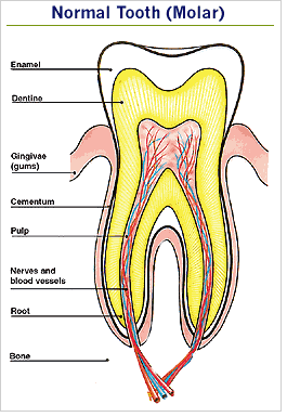
Enamel The hard outer layer of the crown. Enamel is the hardest substance in the body.
Dentine Not as hard as enamel, forms the bulk of the tooth and can be sensitive if the protection of the enamel is lost.
Pulp Soft tissue containing the blood and nerve supply to the tooth. The pulp extends from the crown to the tip of the root.
Cementum The layer of bone-like tissue covering the root. It is not as hard as enamel.
Structures around the tooth
Periodontal ligament: Made up of thousands of fibres which fasten the cementum to the bony socket. These fibres anchor the tooth to the jaw bone and act as shock absorbers for the tooth which is subjected to heavy forces during chewing.
Oral Mucosa: This is the term ussed to describe the moist tissue that lines the mouth.
Gingivae (gums): Soft tissue that immediately surrounds the teeth and bone. It protects the bone and the roots of the teeth and provides an easily lubricated surface.
Bone: Provides a socket to surround and support the roots of the teeth.
Nerves and blood supply: Each tooth and periodontal ligament has a nerve supply and the teeth are sensitive to a wide variety of stimuli. The blood supply is necessary to maintain the vitality of the tooth.